Continue
Cone-beam computed tomography systems are radiographic systems used by dental professionals to analyze and reconstruct 3D images of a patient’s teeth, jaws and surrounding anatomy. The information obtained by means of CBCT imaging is useful in both diagnosis and precise treatment planning when two-dimensional diagnostic films are insufficient. Dental CBCT is useful for multiple types of analyses as well as the assessment of maxillofacial disorders or pathology. It is also most useful in surgical planning, including the accurate placement of dental implants.
Once the decision has been made to pursue orthodontic treatment, the next step is scheduling an appointment with our office to take diagnostic records and develop a precise plan of care. One of the most valuable tools, we use to analyze facial growth and position of the jaws and teeth is a “ceph” or cephalometric x-ray.
A cephalometric film offers a complete and detailed profile view of the face, jaws, and teeth. This information not only shows specific facial growth patterns and the skeletal relationship between the upper and lower jaw, but it also shows the angle of the front teeth and provides a look at the TMJ (temporomandibular joint), and airway. By analyzing the results of this image in conjunction with the information provided by study models of the teeth, facial photographs, a panorex film, and relevant patient history, a precise diagnosis of the type of malocclusion can be made and the best treatment plan developed.
It’s easy, quick, and comfortable to have a cephalometric film taken. All that is required is to stand as positioned with the jaws closed for a brief moment as our digital radiographic system obtains the image. Because we use the most advanced digital technology, patients are exposed to the least amount of x-ray radiation. Once the image gets taken, the information can be analyzed, shared with the patient or any other treating specialists, and stored for mid-treatment or post-treatment comparisons.
Cephalometric films are not just useful in orthodontic case planning, but they can also provide information in the areas of sleep apnea treatment and implant dentistry.
At G’s Dental Studio, we use the most advanced technology and treatment methods to provide our patients with the highest quality of care!
Having a dental lab on premises enables our office to provide the highest quality services and the most convenient options in care. With a full-service laboratory on the premises, we can customize a comprehensive range of dental restorations and prostheses, including inlays/onlays, veneers, crowns, bridges, partial or full dentures, and other appliances. By working hand-in-hand with our onsite dental lab, we’re able to craft restorations that blend seamlessly with your smile while achieving healthy, functional, and long-lasting results of care. And, if you happen to damage or lose a dental restoration or appliance, you can rely on our office and in-house lab for prompt replacements or repairs.
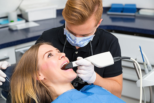
By using intra-oral optical scanning devices, the need for patients to have a messy conventional dental impression taken of their teeth is eliminated. Digital optical impressions enable the dentist to more efficiently and effectively obtain an accurate computer generated representation of their patient’s teeth along with the surrounding tissues. In addition to being much more comfortable for the patient, a digital impression eliminates the need for the dentist to either create a stone model from the impression, or to mail the impression directly to the laboratory for any type of work to be done. Digital impression information is transmitted electronically, significantly reducing the turnaround time of any needed outside laboratory work. Digital impressions are also integral to systems that produce same day, in-office ceramic restorations.
Digital radiography utilizes computer technology and digital sensors for the acquisition, viewing, storage, and sharing of radiographic images. It offers several advantages over the older traditional film based methods of taking x-rays. The most significant of these advantages is that digital radiography reduces a patient’s exposure to radiation. Other benefits are that images can be viewed instantly after being taken, can be seen simultaneously as needed by multiple practitioners, and can be easily shared with other offices. Digital x-rays are also safer for the environment as they do not require any chemicals or paper to develop.
An electronic pad, known as a sensor is used instead of film to acquire a digital image. After the image is taken, it goes directly into the patient’s file on the computer. Once it is stored on the computer, it can be easily viewed on a screen, shared, or printed out.
One of the significant advances in modern dentistry has been the development of dental laser technology. Today, dental lasers are being increasingly used to treat tooth decay, periodontal disease, perform biopsies or the removal of oral lesions, to cure restorative (filling) materials, as well as to activate in-office teeth whitening systems.
Dental lasers combine laser energy with water and air to safely cut and shape target soft or hard tissues in the mouth. Laser energy precisely cuts through tooth structure by exciting the water molecules in the tooth. It operates without direct contact to the tooth without heat, vibration, or pressure thereby minimizing the discomfort of the procedure and the need for dental anesthesia. In addition dental lasers can reduce anxiety for patients fearful of dental work, minimize post-operative bleeding and swelling, and preserve healthy tooth structure during the removal of decay.
While dental lasers may be an excellent treatment option in some situations, they cannot be used for every dental procedure.

CEREC® offers convenient, faster and more comfortable care to provide a range of high quality same day restorations including ceramic crowns, inlays and onlays.
In just one appointment you can have a naturally beautiful restoration to enhance the beauty and function of your smile. With CEREC® there is no need for multiple office visits, messy dental impressions, a temporary crown, or waiting weeks for your dental work to come back from the laboratory. Your new restoration is designed and customized to the exact specifications of your smile all in the very same day!
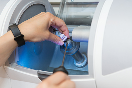
CEREC® is an advanced method of care, which allows for the preparation and placement of same day ceramic restorations. Offering the utmost in terms of quality and convenience, this high-tech system utilizes CAD/CAM technology, or computer aided design and computer aided manufacturing to make every step of the restoration process more patient-friendly and precise. Using an ergonomically designed intraoral scanning wand in place of a conventional dental impression, the dentist is able to capture detailed 3D images of the prepared tooth and surrounding dentition. These images are then integrated by an advanced computer software program to produce an accurate model and plan a precise fitting, functional, and cosmetically pleasing restoration. As soon the restoration is designed and approved by the dentist, the information is sent wirelessly to a chairside milling machine where a ceramic restoration is fabricated as the patient waits.

A CEREC® single visit crown offers much more than a convenient approach to getting a dental crown. It is a high quality, naturally beautiful, extremely durable and long-lasting dental restoration!
At the pinnacle of advanced healthcare technology, the E4D CAD/CAM restorative systems and software solutions enable dental practices to provide their patients with “same day” in-office ceramic restorations. With E4D technology your dentist can take digital impressions, create virtual models, and precisely mill the highest quality ceramic restorations all on the very same day. Using this advanced approach to care eliminates the need for messy impressions, the inconvenience of multiple dental appointments and the extra wait required with outside laboratory-fabricated restorations.